Barrett's Esophagus
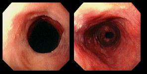
57 year-old man with longstanding symptoms of gastroesophageal reflux disease. Squamocolumnar junction (left) was located 15 cm above the esophagogastric junction. View on the right shows the long segment of distal esophagus covered with columnar epithelium.
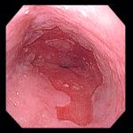
Left: 47 year-old woman with chronic pyrosis (heartburn). Endoscopy demonstrated an irregular squamocolumnar junction, with bands of metaplastic epithelium extending proximally.
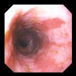
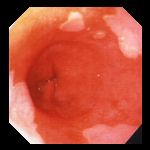
Center: 73 year-old woman with pyrosis and dysphagia (difficulty swallowing). Squamocolumnar junction shown here was located 10 cm above the junction of the stomach and esophagus.
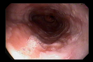
Right: 85 year-old man with chronic gastroesophageal reflux disease, in whom the squamocolumnar junction has migrated upward from the esophagogastric junction in an asymmetric fashion.
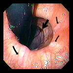
Left: Retroflexed view, looking upward at the junction of the stomach and esophagus in a 72 year-old man with Barrett's. The large arrow indicates the squamocolumnar junction, located several centimeters upward in the esophagus. The smaller arrows indicate islands of squamous mucosa left behind in the segment of columnar epithelium.
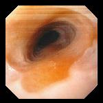
Center: Segment of Barrett's with residual islands of squamous epithelium.
Right: 71 year-old woman with chronic reflux symptoms. Endoscopy revealed asymmetric proximal migration of the squamocolumnar junction, with small islands of squamous mucosa within the segment of columnar epithelium.
56 year-old man with chronic GERD undergoing endoscopy to screen for Barrett's. Endoscopy revealed an approximately 12 cm segment of Barrett's epithelium.
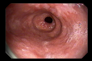
Left: View of the upper extent of Barrett's at the squamocolumnar junction ("Z line") located 30 cm distal to the incisors. Note the irregular contour.
Right: The lower portion of the segment of Barrett's epithelium with the esophagogastric junction, located 42 cm distal to the incisors, visible. There are "tongues" of squamous epithelium at the top of the imaging extending downward into the segment of intestinal metaplasia.
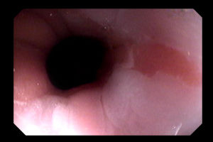

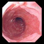
Left: 53 year-old woman with known Barrett's disease, undergoing endoscopy for dysplasia screening. Exam revealed a small salmon-colored "tongue" of columnar epithelium extending upward into normal silver-colored esophageal squamous epithelium. Biopsies did not find dysplasia.
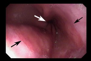
Center and Right: 68 year-old woman previously diagnosed with Barrett's. Photos show proximal migration of the squamocolumnar junction (black arrows) with the GE junction (white arrow) visible distally. No dysplasia was found on biopsy.
Endoscopic images Copyright © Atlanta South Gastroenterology, P.C. All rights reserved.
Logo is Registered Trademark ® of Atlanta South Gastroenterology, P.C.
This site is presented for educational and general informational purposes only. It does not purport to offer medical advice for any specific medical condition or individual patient. We regret that we cannot provide individualized medical advice online, either via this web site or via email. Please refer to our "Notable Web Sites" section, which offers links to several excellent online sources of additional medical information.