Candida Esophagitis

Left: Whitish yeast colonies which may become confluent, and which may be associated with ulceration.
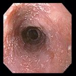
Right: 63 year-old man with gastric carcinoma on the left; 40 year-old woman with recurrent Candida esophagitis in whom an immune status evaluation was negative.
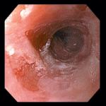
52 year-old woman receiving corticosteroids for lung disease, presenting with dysphagia. Typical whitish macules
were seen proximally (left), with more severe inflammation and ulceration
distally (right).
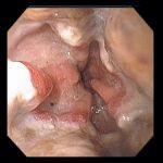
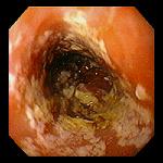
73 year-old woman with dysphagia and odynophagia. Confluent exudate with discoloration was noted in the proximal
esophagus (left), with dense exudate extending to the squamocolumnar junction
distally (right).
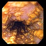
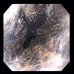
37 year-old man with HIV undergoing endoscopy for abdominal pain. Endoscopy revealed Candida esophagitis.
Endoscopic images Copyright © Atlanta South Gastroenterology, P.C. All rights reserved.
Logo is Registered Trademark ® of Atlanta South Gastroenterology, P.C.
This site is presented for educational and general informational purposes only. It does not purport to offer medical advice for any specific medical condition or individual patient. We regret that we cannot provide individualized medical advice online, either via this web site or via email. Please refer to our "Notable Web Sites" section, which offers links to several excellent online sources of additional medical information.