Mallory-Weiss
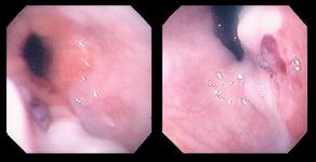
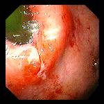
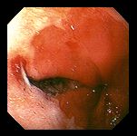
Left and Right: A linear mucosal laceration caused by the force of vomiting, generally at or just below the esophagogastric junction, resulting in bleeding. Seen here is an elliptical-shaped mucosal tear which has stopped bleeding, from the forward view (on the left) and from the retroflexed position (on the right).
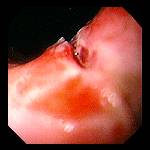
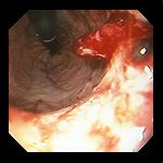
Left: 66 year-old women with a self-induced, intraprocedural Mallory-Weiss caused by retching during the exam. Bleeding was self-limited; shown here are fresh blood coating mucosa, and the clot forming at the point of bleeding just below the esophagogastric junction, seen at retroflexion.
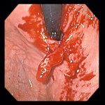
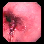
Center: 44 year-old man with hematemesis. endoscopy revealed a thin, linear tear beginning just above the squamocolumnar junction and extending proximally, from which there was active bleeding.
Right: Iatrogenic Mallory-Weiss which developed during balloon dilation of a stricture in the distal esophagus. Despite the width of the mucosal tear, there were no adverse sequellae, and the patient's dysphagia was significantly improved.
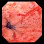
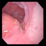
Left: This thin linear mucosal tear is seen on a retroflexed view, looking back up at the esophagogastric junction.
Center: Close up of the mucosal tear from the photo at left, showing the separation of the mucosa.
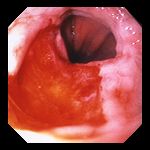
Right: Deep, wide mucosal tear beginning just above the squamocolumnar junction, extending rightward in this photo, in a 51 year-old man presenting with hematemesis (vomiting blood). Bleeding was self-limited once the vomiting was brought under control.
Left: 41 year-old man with hematemesis (vomiting blood). Endoscopy demonstrated a linear mucosal tear extending through the squamocolumnar junction, with an adherent clot.
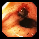
Center: Fresh clot on a mucosal tear in a 24 year-old man who presented with severe anemia and hematemesis. Viewed at retroflexion.
Right: Adherent clot and oozing of blood from a mucosal tear in an 89 year-old woman with hematemesis and a concurrently bleeding duodenal ulcer.
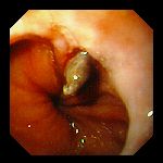
Left: Linear mucosal tear at the esophagogastric junction in a 69 year-old man who presented with hematemesis (vomiting blood).
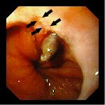
Center: 42 year-old woman presenting with hematemesis. Endoscopy demonstrated a linear ulcer (see arrows at right), at the distal (lower) end of which was an adherent "sentinel" clot, denoting the site of recent bleeding. The lesion spans the junction of squamous and columnar mucosa at the esophagagastric junction; the clot resides on the gastric side.
Endoscopic images Copyright © Atlanta South Gastroenterology, P.C. All rights reserved.
Logo is Registered Trademark ® of Atlanta South Gastroenterology, P.C.
This site is presented for educational and general informational purposes only. It does not purport to offer medical advice for any specific medical condition or individual patient. We regret that we cannot provide individualized medical advice online, either via this web site or via email. Please refer to our "Notable Web Sites" section, which offers links to several excellent online sources of additional medical information.