Gastric Varices
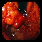
Left: 31 year-old man with end-stage liver disease secondary to alcoholic cirrhosis, with a recurrent episode of bleeding from gastric and esophageal varices. Seen on retroflexion are pendulous varices in the gastric cardia and fundus, covered with fresh-appearing blood.
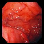
Right: 78 year-old man with colonic carcinoma metastatic to liver, causing portal hypertension. Serpiginous varices course through the gastric fundus.
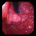
Left: 65 year-old man with cryptogenic cirrhosis presented with painless melena, and was found to have gastric and esophageal varices. Polyp-like varices are shown here in the gastric cardia, seen on retroflexion of the endoscope.
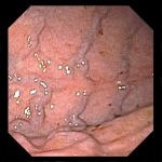
Right: Linear varices in the gastric body of a 69 year-old woman with abdominal pain and weight loss. There were no varices in the esophagus or gastric cardia. The woman was ultimately found to have pancreatic carcinoma, and was suspected to have splenic vein thrombosis.
Endoscopic images Copyright © Atlanta South Gastroenterology, P.C. All rights reserved.
Logo is Registered Trademark ® of Atlanta South Gastroenterology, P.C.
This site is presented for educational and general informational purposes only. It does not purport to offer medical advice for any specific medical condition or individual patient. We regret that we cannot provide individualized medical advice online, either via this web site or via email. Please refer to our "Notable Web Sites" section, which offers links to several excellent online sources of additional medical information.