Gastric Adenocarcinoma
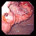
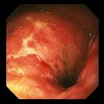
Left: 71 year-old man with dysphagia. Endoscopy revealed an ulcerated mass in the gastric cardia, seen here at retroflexion. Esophageal symptoms were caused by upward extension of the tumor into distal esophagus
(center).
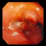
Right: Ulcerated neoplastic mass arising in the gastric cardia of a 34 year-old man. Biopsy revealed the lesion to be adenocarcinoma.
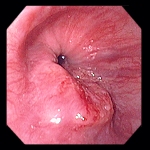
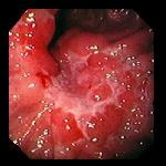
Left: 46 year-old man with no prior gastrointestinal symptoms, presented with five days of epigastric pain. Initial studies revealed iron-deficiency anemia and blood in the stool. Endoscopy demonstrated this lesion on the lesser curvature which appeared to be edematous folds with a central ulceration, but which on biopsy proved to be a poorly differentiated adenocarcinoma, signet ring cell type.
Center: 50 year-old man who had undergone seemingly successful resection of adenocarcinoma involving the gastric antrum, now undergoing endoscopy for routine postoperative screening several months later. Endoscopy revealed a poorly distensible, ulcerated distal gastric remnant. Biopsies confirmed the suspicion of recurrent carcinoma.
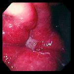
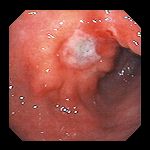
Right: 87 year-old woman was found to be anemic and to have occult blood in the stool; she had no gastrointestinal symptoms of any kind. Endoscopy revealed this ulcerated, sessile, polypoid mass which proved to be adenocarcinoma on biopsy.

Left: 82 year-old woman who presented with early satiety and postprandial vomiting, suggestive of gastric outlet obstruction, along with weight loss and anemia. Endoscopy demonstrated an ulcerated mass with prominent folds, which did not obstruct the gastric outlet. The lesion was an adenocarcinoma of the signet ring cell type.
Center: 54 year-old woman with abdominal pain. Biopsies of this ulcer, with raised margins, revealed poorly differentiated adenocarcinoma, signet-ring type.
Right: 70 year-old man with melena as his only presenting symptom; no nausea, vomiting, early satiety or pain. Endoscopy revealed a partially obstructing adenocarcinoma, seen here from the antrum. The tunor extended into the second portion of the duodenum.
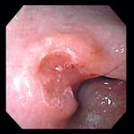
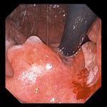
51 year-old woman who had undergone EGD one month previously for symptoms of abdominal pain. At that time, endoscopy revealed nonspecific-appearing antral gastritis, biopsies of which
revealed gastritis with atypical cells. At repeat endoscopy, there was now a raised antral process with a central depression, as shown in this image. Repeat biopsies now revealed
invasive poorly differentiated adenocarcinoma.
Endoscopic images Copyright © Atlanta South Gastroenterology, P.C. All rights reserved.
Logo is Registered Trademark ® of Atlanta South Gastroenterology, P.C.
This site is presented for educational and general informational purposes only. It does not purport to offer medical advice for any specific medical condition or individual patient. We regret that we cannot provide individualized medical advice online, either via this web site or via email. Please refer to our "Notable Web Sites" section, which offers links to several excellent online sources of additional medical information.