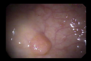
Traditional Serrated Adenoma
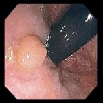
82 year-old man with a bland rectal polyp, visible only from the retroflexed view. Histologically the lesion was a mixture of hyperplastic and adenomatous elements, hence the designation of traditional serrated adenoma.
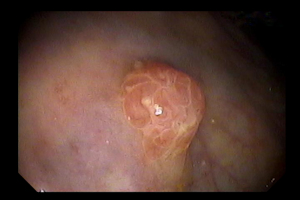
Left: 76 year-old man with occult gastrointestinal bleeding. A 6 mm sessile traditional serrated adenoma was found in the rectum.
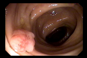
Right: 67 year-old woman undergoing colonoscopy as a follow up of recent diverticulitis. This 1 cm sessile polyp in the sigmoid colon was a traditional serrated adenoma.
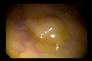
Left: 73 year-old man seen for average risk colon screening. Colonoscopy revealed this 8 mm sessile traditional serrated adenoma in the sigmoid colon.
Right: 55 year-old woman with chronic constipation undergoing colonoscopy for average risk colon screening. A 1 cm sessile rectal polyp proved to be a traditional serrated adenoma.
See also Sessile Serrated Adenoma
Endoscopic images Copyright © Atlanta South Gastroenterology, P.C. All rights reserved.
Logo is Registered Trademark ® of Atlanta South Gastroenterology, P.C.
This site is presented for educational and general informational purposes only. It does not purport to offer medical advice for any specific medical condition or individual patient. We regret that we cannot provide individualized medical advice online, either via this web site or via email. Please refer to our "Notable Web Sites" section, which offers links to several excellent online sources of additional medical information.