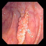
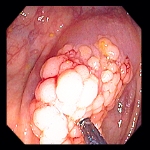
Polypectomy - Pedunculated Polyp
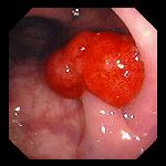
 Left: 1.5 cm bi-lobed benign tubular adenoma on a stalk.
Right: Placement of a snare wire over the head of the polyp.
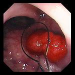
 Left: Cautery is applied to the wire loop which has been tightened around the stalk of the polyp.
Right: Polypectomy site; excised polyp in the background, waiting to be retrieved.
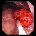
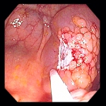
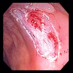
Polypectomy - Sessile Polyp
 Left: Sessile villous adenoma near the cecum of a 72 year-old man undergoing routine screening colonoscopy.
Right: Polyp is injected submucosally with saline to elevate the lesion away from the colonic wall, and in so doing, determine that the lesion is not invasive into the colonic wall.
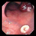
 Left: After submucosal injection, the polyp is excised piecemeal using a snare. Shown here after removal of first fragment.
Right: Polypectomy site at completion of polyp resection.
Endoscopic images Copyright © Atlanta South Gastroenterology, P.C. All rights reserved.
Logo is Registered Trademark ® of Atlanta South Gastroenterology, P.C.
This site is presented for educational and general informational purposes only. It does not purport to offer medical advice for any specific medical condition or individual patient. We regret that we cannot provide individualized medical advice online, either via this web site or via email. Please refer to our "Notable Web Sites" section, which offers links to several excellent online sources of additional medical information.